English
 norsk
norskBlar i forfatter "Ahluwalia, Balpreet Singh"
Viser treff 1-20 av 84
-
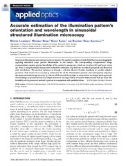
Accurate estimation of the illumination pattern’s orientation and wavelength in sinusoidal structured illumination microscopy
(Journal article; Tidsskriftartikkel; Peer reviewed, 2018-01-05)Structured illumination microscopy is able to improve the spatial resolution of wide-field fluorescence imaging by applying sinusoidal stripe pattern illumination to the sample. The corresponding computational image reconstruction requires precise knowledge of the pattern’s parameters, which are its phase (φ) and wave vector (<b>p</b>). Here, a computationally inexpensive method for estimation of ... -
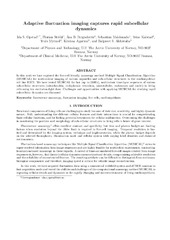
Adaptive fluctuation imaging captures rapid subcellular dynamics
(Journal article; Tidsskriftartikkel, 2019-07-22)In this work we have explored the live-cell friendly nanoscopy method Multiple Signal Classification Algorithm (MUSICAL) for multi-colour imaging of various organelles and sub-cellular structures in the cardiomyoblast cell line H2c9. We have tested MUSICAL for fast (up to 230Hz), multi-colour time-lapse sequences of various sub-cellular structures (mitochondria, endoplasmic reticulum, microtubules, ... -
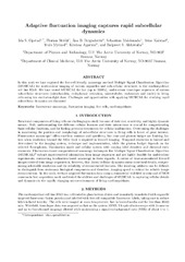
Adaptive fluctuation imaging captures rapid subcellular dynamics
(Journal article; Tidsskriftartikkel; Peer reviewed, 2019-07-22)In this work we have explored the live-cell friendly nanoscopy method Multiple Signal Classification Algorithm (MUSICAL) for multi-colour imaging of various organelles and sub-cellular structures in the cardiomyoblast cell line H2c9. We have tested MUSICAL for fast (up to 230Hz), multi-colour time-lapse sequences of various sub-cellular structures (mitochondria, endoplasmic reticulum, microtubules, ... -
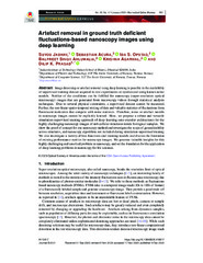
Artefact removal in ground truth deficient fluctuations-based nanoscopy images using deep learning
(Journal article; Tidsskriftartikkel; Peer reviewed, 2020-12-08)Image denoising or artefact removal using deep learning is possible in the availability of supervised training dataset acquired in real experiments or synthesized using known noise models. Neither of the conditions can be fulfilled for nanoscopy (super-resolution optical microscopy) images that are generated from microscopy videos through statistical analysis techniques. Due to several physical ... -
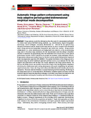
Automatic fringe pattern enhancement using truly adaptive period-guided bidimensional empirical mode decomposition
(Journal article; Tidsskriftartikkel; Peer reviewed, 2020-02-19)Fringe patterns encode the information about the result of a measurement performed via widely used optical full-field testing methods, e.g., interferometry, digital holographic microscopy, moiré techniques, structured illumination etc. Affected by the optical setup, changing environment and the sample itself fringe patterns are often corrupted with substantial noise, strong and uneven background ... -
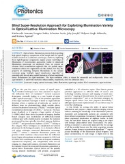
Blind Super-Resolution Approach for Exploiting Illumination Variety in Optical-Lattice Illumination Microscopy
(Journal article; Tidsskriftartikkel; Peer reviewed, 2021-08-19)Optical-lattice illumination patterns help in pushing high spatial frequency components of the sample into the optical transfer function of a collection microscope. However, exploiting these high-frequency components require precise knowledge of illumination if reconstruction approaches similar to structured illumination microscopy are employed. Here, we present an alternate blind reconstruction ... -
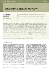
Characterization of a waveguide Mach-Zehnder interferometer using PDMS as a cover layer
(Journal article; Tidsskriftartikkel; Peer reviewed, 2015)A Mach-Zehnder interferometer made with shallow rib waveguides is studied experimentally and using simulations. The rib-height giving single-mode guidance is found as function of core thickness and polarization. Devices have been made using shallow rib waveguides (5 nm rib height) in silicon nitride. The sensitivity and the limit of detection (LOD) is studied experimentally regarding the length of ... -
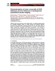
Characterization of color cross-talk of CCD detectors and its influence in multispectral quantitative phase imaging
(Journal article; Tidsskriftartikkel; Peer reviewed, 2019-02-08)Multi-spectral quantitative phase imaging (QPI) is an emerging imaging modality for wavelength dependent studies of several biological and industrial specimens. Simultaneous multi-spectral QPI is generally performed with color CCD cameras. Here, we present a new approach for accurately measuring the color crosstalk of 2D area detectors, without needing prior information about camera specifications. ... -
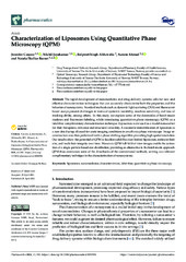
Characterization of Liposomes Using Quantitative Phase Microscopy (QPM)
(Journal article; Tidsskriftartikkel; Peer reviewed, 2021-04-21)The rapid development of nanomedicine and drug delivery systems calls for new and effective characterization techniques that can accurately characterize both the properties and the behavior of nanosystems. Standard methods such as dynamic light scattering (DLS) and fluorescent-based assays present challenges in terms of system’s instability, machine sensitivity, and loss of tracking ability, among ... -
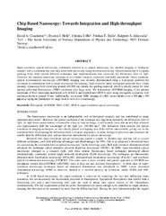
Chip Based Nanoscopy: Towards Integration and High-throughput Imaging
(Journal article; Tidsskriftartikkel; Peer reviewed, 2017)Super-resolution optical microscopy, commonly referred to as optical nanoscopy, has enabled imaging of biological samples with a resolution that was only achievable previously using electron microscopy. Optical nanoscopy is a rapidly growing field, with several different techniques and implementations that overcome the diffraction limit of light. However, the common nanoscope continues to be a rather ... -
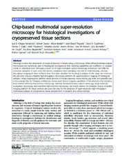
Chip-based multimodal super-resolution microscopy for histological investigations of cryopreserved tissue sections
(Journal article; Tidsskriftartikkel; Peer reviewed, 2022-02-24)Histology involves the observation of structural features in tissues using a microscope. While diffraction-limited optical microscopes are commonly used in histological investigations, their resolving capabilities are insufficient to visualize details at subcellular level. Although a novel set of super-resolution optical microscopy techniques can fulfill the resolution demands in such cases, the ... -
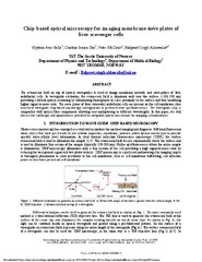
Chip-based optical microscopy for imaging membrane sieve plates of liver scavenger cells
(Journal article; Tidsskriftartikkel; Peer reviewed, 2015-08-26)The evanescent field on top of optical waveguides is used to image membrane network and sieve-plates of liver endothelial cells. In waveguide excitation, the evanescent field is dominant only near the surface (~100-150 nm) providing a default optical sectioning by illuminating fluorophores in close proximity to the surface and thus benefiting higher signal-to-noise ratio. The sieve plates of liver ... -
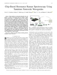
Chip-Based Resonance Raman Spectroscopy Using Tantalum Pentoxide Waveguides
(Journal article; Tidsskriftartikkel; Peer reviewed, 2019-05-08)Blood analysis is an important diagnostic tool, as it provides a wealth of information about the patient's health. Raman spectroscopy is a promising tool for blood analysis, but widespread clinical application is limited by its low signal strength, as well as complex and costly instrumentation. The growing field of waveguide-based Raman spectroscopy tries to solve these challenges by working toward ... -
Chip-based wide field-of-view nanoscopy
(Journal article; Tidsskriftartikkel; Peer reviewed, 2017-04-24)Present optical nanoscopy techniques use a complex microscope for imaging and a simple glass slide to hold the sample. Here, we demonstrate the inverse: the use of a complex, but mass-producible optical chip, which hosts the sample and provides a waveguide for the illumination source, and a standard low-cost microscope to acquire super-resolved images via two different approaches. Waveguides composed ... -
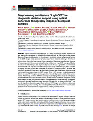
Deep learning architecture “LightOCT” for diagnostic decision support using optical coherence tomography images of biological samples
(Journal article; Tidsskriftartikkel; Peer reviewed, 2020-08-13)Optical coherence tomography (OCT) is being increasingly adopted as a label-free and non-invasive technique for biomedical applications such as cancer and ocular disease diagnosis. Diagnostic information for these tissues is manifest in textural and geometric features of the OCT images, which are used by human expertise to interpret and triage. However, it suffers delays due to the long process of ... -
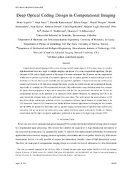
Deep Optical Coding Design in Computational Imaging: A data-driven framework
(Journal article; Tidsskriftartikkel; Peer reviewed, 2023-02-27)Computational optical imaging (COI) systems leverage optical coding elements (CEs) in their setups to encode a high-dimensional scene in a single or in multiple snapshots and decode it by using computational algorithms. The performance of COI systems highly depends on the design of its main components: the CE pattern and the computational method used to perform a given task. Conventional approaches ... -
Demystifying speckle field interference microscopy
(Journal article; Tidsskriftartikkel; Peer reviewed, 2022-06-27)Dynamic speckle illumination (DSI) has recently attracted strong attention in the feld of biomedical imaging as it pushes the limits of interference microscopy (IM) in terms of phase sensitivity, and spatial and temporal resolution compared to conventional light source illumination. To date, despite conspicuous advantages, it has not been extensively implemented in the feld of phase imaging due ... -
Deriving high contrast fluorescence microscopy images through low contrast noisy image stacks
(Journal article; Tidsskriftartikkel; Peer reviewed, 2021-08-11)Contrast in fluorescence microscopy images allows for the differentiation between different structures by their difference in intensities. However, factors such as point-spread function and noise may reduce it, affecting its interpretability. We identified that fluctuation of emitters in a stack of images can be exploited to achieve increased contrast when compared to the average and Richardson-Lucy ... -
Effect of caffeine and other xanthines on liver sinusoidal endothelial cell ultrastructure
(Journal article; Tidsskriftartikkel; Peer reviewed, 2023-08-17)Xanthines such as caffeine and theobromine are among the most consumed psychoactive stimulants in the world, either as natural components of cofee, tea and chocolate, or as added ingredients. The present study assessed if xanthines affect liver sinusoidal endothelial cells (LSEC). Cultured primary rat LSEC were challenged with xanthines at concentrations typically obtained from normal consumption of ... -
Effect on the longitudinal coherence properties of a pseudothermal light source as a function of source size and temporal coherence
(Journal article; Tidsskriftartikkel; Peer reviewed, 2019-04-01)In the present Letter, a synthesized pseudothermal light source having high temporal coherence (TC) and low spatial coherence (SC) properties is used. The longitudinal coherence (LC) properties of the spatially extended monochromatic light source are systematically studied. The pseudothermal light source is generated from two different monochromatic laser sources: He–Ne (at 632 nm) and DPSS (at 532 ...